

Piezo Objective Scanner Makes Cryo-electron Microscope Atoms Visible Clearly
In the world of microstructure, to clearly observe and conveniently manipulate individual atoms, it is often necessary to rely on a special method - cryo-electron microscopy. It can cool atoms to a low-temperature state and fix them in an optical lattice, thereby achieving imaging and manipulation of individual atoms. One of the core challenges of this technology is that it can achieve extremely high imaging accuracy even in the state of atomic freezing.
I. Cryo-Electron Microscope
An Eye to Peek into Atomic Structure
Cryo-electron microscopy (cryo-EM) is an advanced technology that uses electron microscopy to observe samples such as biological macromolecules, viruses, and cells. Its core advantage lies in its ability to resolve the high-resolution 3D structure of samples in a state close to their natural state. It can image biomolecules such as proteins, enzymes and viruses with atomic-level precision, greatly enhancing the clarity of biomolecular imaging and completely transforming the research model in fields such as structural biology.
(Note: Image from online resources)
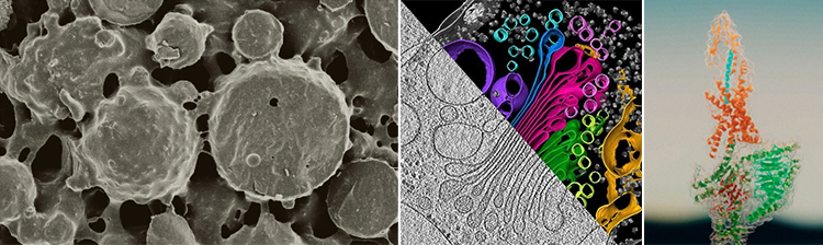
When observing biological samples with a traditional electron microscope, the samples need to be processed, which can easily damage the natural structure of the samples. Cryo-electron microscopy is different, it can rapidly cool the sample to a low-temperature state through rapid freezing technology, which can prevent the crystallization of water molecules in the sample, thereby preserving the near-physiological state and complete natural structure of the sample (such as the folded state of proteins, the morphology of viruses, etc.), making it easy for 3D imaging observation.
(Note: Image from online resources)
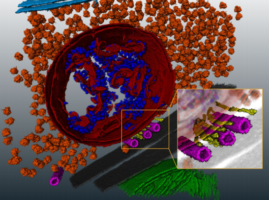
The core value of cryo-electron microscopy lies in its ability to achieve true, high-definition and extensive three-dimensional structural analysis of biomolecules in a non-destructive and non-restrictive manner. Therefore, it is hailed as a revolutionary tool in the field of structural biology and has driven breakthroughs in multiple disciplines. Its application scope is extremely wide: it can resolve everything from small molecules such as proteins, nucleic acids, and complexes to large structures like viruses, ribosomes, and even local parts of cells. Cryo-electron microscopy is particularly adept at analyzing complex systems that are difficult to handle with traditional techniques, such as membrane proteins and dynamic molecular complexes.
(Note: Image from online resources)
II. CoreMorrow
Piezoelectric Objective Scanner
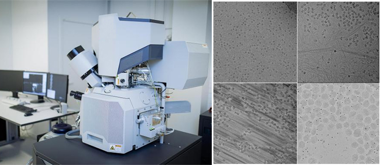
Inside a cryo-electron microscope, the positioning accuracy of the objective lens directly determines the imaging quality. With the continuous advancement of technology, researchers have been constantly improving their exploration of the structure of biological cells. To solve the problem of the biological complexity of proteins, at present, it is necessary to observe more clearly the arrangement of individual atoms in the structure of biomolecules, such as: When observing the changes in cells during viral infection, the morphology of the virus at different development stages can be observed through cryo-electron microscopy. When the positioning accuracy needs to reach the nanometer level, it is the stage for piezo objective positioning technology to shine.
(Note: Image from online resources)
The objective scanner is one of the core guarantees for achieving atomic-level resolution in cryo-electron microscopes. Its performance simultaneously meet four core requirements: environmental adaptability, ultra-high precision and repeatability, rapid dynamic response, and low interference. These advantages jointly ensure that cryo-electron microscopy can stably and efficiently collect high-quality images, providing a reliable data foundation for the 3D structure analysis of biological macromolecules. CoreMorrow piezo technology precisely meet these essential needs:
Precision matching: The piezoelectric ceramic drive and closed-loop feedback system can achieve ultra-precise displacement control, ensuring zero deviation in imaging.
Fast response: The piezoelectric objective positioning device has an extremely fast response time, meeting the follow-up response requirements for multi-dimensional imaging of molecular structures.
Stable performance: Piezoelectric ceramic drive has no electromagnetic interference, and the integrated design reduces the impact of vibration.
Environmental compatibility: Made of low-temperature resistant materials, it can maintain stable mechanical properties even in low-temperature environments and is compatible with high vacuum environments.
P72 Piezo Objective Scanner
P72 piezoelectric objective scanner is a Z-axis motion piezo positioner specially designed for objective lens focusing microscopy. The objective positioner is loaded with a microscopic detection/measurement or observation device to improve accuracy. It can be equipped with a load lens for precise positioning adjustment to improve focusing accuracy and can be used in conjunction with various high-resolution microscopes.
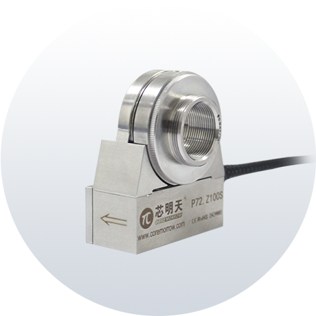
Characteristics
• Travel to 100μm
• Load capacity to 0.4kg
• Millisecond response time
• Closed loop for high repeatability
• Open-loop and closed-loop options are available
Typical Model
|
Model |
P72.Z100S |
|---|---|
|
Active axis |
Z |
|
Drive control |
1 driving channel, 1 sensing channel |
|
Nominal travel range (0~120V) |
80µm |
|
Max. travel range(0~150V) |
100µm |
|
Sensor |
SGS |
|
Resolution |
3nm |
|
Closed-loop linearity |
0.1%F.S. |
|
Closed-loop Repeatability |
0.05%F.S. |
|
Push/pull force |
110N/15N |
|
Stiffness |
1.4N/µm |
|
Unloaded resonant frequency |
350Hz |
|
Loaded resonant frequency |
150Hz@200g |
|
Closed-loop unloaded step time |
10ms |
|
Closed-loop operating frequency (-3dB) |
70Hz@100g |
|
Load capacity |
0.2Kg |
|
El. capacitance |
3.6μF |
|
Material |
Steel |
|
Mass |
150g |
P75 Piezo Objective Scanner
P75 large-stroke piezo objective scanner can achieve a displacement of 250μm in Z-axis, with an load capacity of 0.2kg. It adopts a parallel guiding mechanism design with a flexible hinge without hysteresis, featuring no friction, good linearity, high closed-loop positioning accuracy, small objective lens compensation, and ultra-high focusing stability. The detachable thread adapter design makes it compatible with various models of microscopes.
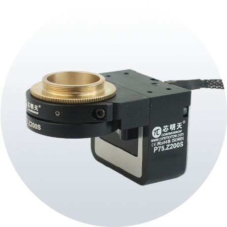
Characteristics
• Travel to 250μm
• Load capacity to 0.2kg
• Detachable thread adapter
• Closed loop for high repeatability
Typical Model
|
Model |
P75.Z200S |
|---|---|
|
Active axis |
Z |
|
Drive control |
1 driving channel, 1 sensing channel |
|
Nominal travel range (0~120V) |
200µm |
|
Max. travel range(0~150V) |
250µm |
|
Sensor |
SGS |
|
Resolution |
6nm |
|
Closed-loop Repeatability |
0.05%F.S. |
|
Push/pull force |
25N/10N |
|
Stiffness |
0.1N/μm |
|
Unloaded resonant frequency |
389Hz |
|
Closed-loop operating frequency (-3dB) |
20Hz |
|
Closed-loop unloaded step time |
25ms@200g |
|
Load capacity |
0.2kg |
|
El. capacitance |
6.5μF |
|
Material |
Steel, Al |
|
Mass |
225g |
1mm version available.
For further details, please call +86-451-86268790, or add WeChat ID: 17051647888.
Sweep, get the latest information on core tomorrow
Address:1F, Building I2, No.191 Xuefu Road, Nangang District, Harbin, China
Tel:0451-86268790
Piezo · Nano · Motion
