

Application of Piezo Technology in Laser Confocal Scanning Microscope
Laser confocal scanning microscope is a technology developed and widely used in the past ten years. It uses a laser as a light source and consists of a confocal imaging scanning system, an electron optical system and an image analysis system. After the beam is focused, it falls on tiny points at different depths of the sample (tissue slab or cell), and moves and scans, so that the image formed by the reflected light at any point in the sample can be accurately received and a signal is generated, and then connected The image analysis system performs analysis processing. It is an advanced cell and molecular biological analysis instrument, which is more and more widely used in biology and medicine, and has become an essential tool for biomedical experimental research.
Note: picture source network
Traditional fluorescence microscopes use fluorescent substances to mark specific structures in cells, the contrast between the image and the background is enhanced, and the light source uses short-wavelength ultraviolet light, which greatly improves the resolution. However, when the observed fluorescent specimen is slightly thicker, an insurmountable disadvantage of traditional fluorescence microscopes is manifested: the fluorescent structures outside the focal plane are blurred and blurred. The reason is that most biological specimens are overlapping structures with different layers, and the changes in the focal plane under ordinary light microscopes will show different morphologies. If the fluorescently labeled structures are distributed at different levels and overlap each other, reflected fluorescence microscopy not only collects light from the focal plane, but also the scattered fluorescence from above or below the focal plane is also received by the objective lens, and the optical resolution of fluorescence microscopy. The rate is thus greatly reduced.
Note: picture source network
On the basis of traditional optical microscope, laser scanning confocal microscope uses laser as light source, adopts conjugate focusing principle and device, and uses computer to perform digital image processing, observation, analysis and output on the observed object. Its feature is that it can perform tomography and imaging on the sample, and conduct non-invasive observation and analysis of the three-dimensional spatial structure of cells. At the same time, using immunofluorescence labeling and ion fluorescence labeling probes, this technology can not only observe fixed cells and tissue sections, but also conduct real-time dynamic observation and detection of the structure, molecules, ions and life activities of living cells. Observing physiological signals such as Ca2+, pH value, membrane potential and other changes in cell morphology and cell morphology, it has become a new generation of powerful research tools in the fields of morphology, molecular and cell biology, neuroscience, pharmacology, genetics, etc. People's understanding of the phenomenon of cell life.
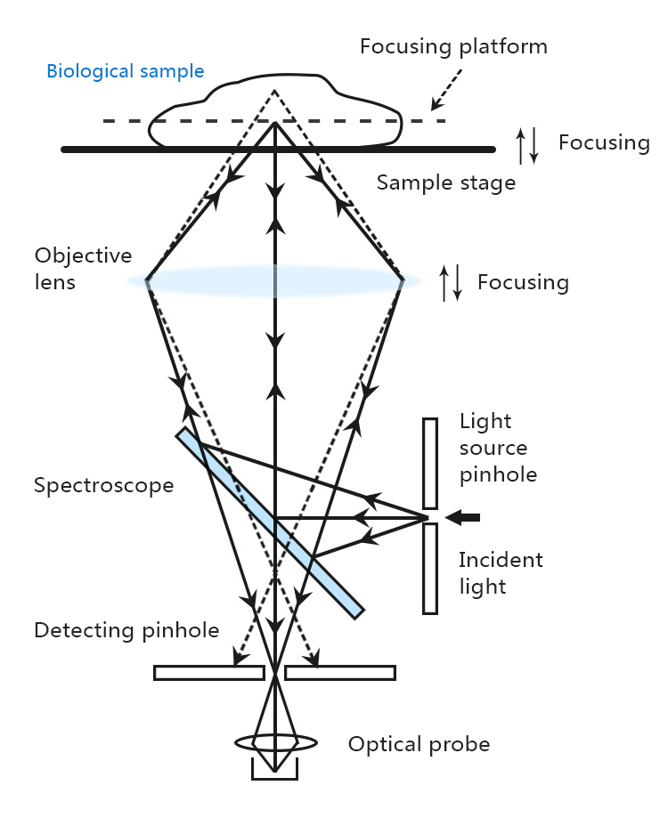
Laser Confocal Scanning Microscopy
CoreMorrow XD781 is a Z-axis motion piezo nanopositioning stage, which is very suitable for laser confocal scanning microscope applications. The high-precision Z-axis displacement movement can adjust the focal plane of the objective lens to nanometer-level precision. It has a very large central through hole that can carry more biological samples, and has a simple box design with narrower edges, which is very easy to integrate.
Features
· Z movement
· Large center through hole
· Nanometer precision
· Small footprint and easy integration
Applications
· Microfocusing/imaging
· Z-direction nanopositioning
· Surface inspection
· Optics
· Micro-nano processing, etc.
Parameters
Model: XD781.100S-B1
Active axis: Z
Travel range: 80μm@120V
Travel range: 100μm@150V
Sensor Type: SGS
Closed/open loop resolution: 3/1nm
Un-load resonance frequency: 300Hz
Loading 100g resonant frequency: 200Hz
Load capacity: 1kg
Capacitance: 5.4μF
Material: Steel, Aluminum
Weight: 875g
CoreMorrow XD730.500 is a piezo objective scanner with large load capacity, large stroke and high precision. In the application of laser confocal scanning microscope, it can move the animal lens to achieve the effect of focal length adjustment.
CoreMorrow piezo objective scanner and the objective lens are connected through an adapter to quickly lock the objective lens in the desired position. A variety of thread options are available, such as M27×0.75, M26×0.75, M26×1/36", M25×0.75, W0.8×1/36", etc., and the thread size can also be customized to facilitate integrated installation with the microscope.

Features
· Z movement
· Large bearing capacity
· Large stroke
· High precision
· Optional closed-loop sensor
Applications
· Focus Microscopy
· laser processing
· Z-direction positioning and scanning
· Surface structure analysis
Parameters
Model: XD730.500
Active axis: Z
Travel range: 400μm@120V
Travel range: 500μm@150V
Sensor Type: SGS
Resolution: 17/5nm
Closed-loop linearity: 0.12%F.S.
Closed loop repeatability: 0.03%F.S.
Unload resonance frequency: 310Hz
Resonance frequency@500g: 155Hz
Capacitance: 30μF
Load capacity: 0.5kg
Material: aluminum, steel
Weight: 590g
- Previous article:Application of Piezo Technology in Interferometry
- Next article:Application of Piezo Nano Position System from CoreMorrow
Sweep, get the latest information on core tomorrow
Address:1F, Building I2, No.191 Xuefu Road, Nangang District, Harbin, China
Tel:0451-86268790
